Figure 6.

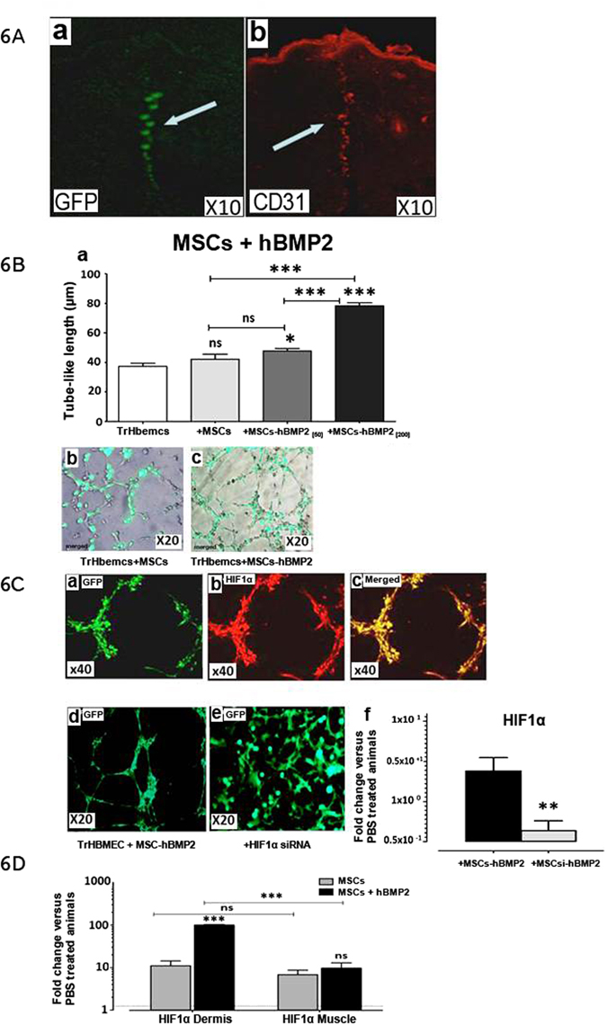
Impact of hBMP-2 addition to MSC therapy on endothelial cell recruitment in vasculogenesis. (A) (a and b) Co-localization of MSCs with CD31 + cells at the site of infusion. (B) (a) Tube length formation, human-endothelial cells (TrHBMEC, white bar) alone and with human MSCs (grey bar), and with the addition of 50 (dark grey bar) or 200 ng/ml hBMP-2 (black bar). (C) Fluorescent microscopy examination of the Matrigel basement membrane matrix from an in vitro tube formation assay with (a–d) endothelial cells (stained with alexa-coupled phalloidin), human-MSCs and hBMP-2 (TrHBMEC + MSCs + hBMP-2), staining with HIF-1 α (b), merged (c), and (e) plus siRNA in MSCs (MSCsi). (f) qRT-PCR measurement of HIF-1α RNA with siRNA (black histogram) or without white histogram. D qRT-PCR measurement of HIF-α RNA in the dermis and muscle with MSCs (grey histogram) or with MSCs plus hBMP-2 black histogram. Results were expressed as the fold change in comparison with un-irradiated (PBS-treated animals). Each group of animals comprised 10 Sprague Dawley rats. Comparison by ANOVA followed by the Mann-Whitney or Dunnett’s test for group pair-wise comparisons. Data represent mean ± SEM, ns: not significant, *p < 0.05, **p < 0.01, ***p < 0.001.
