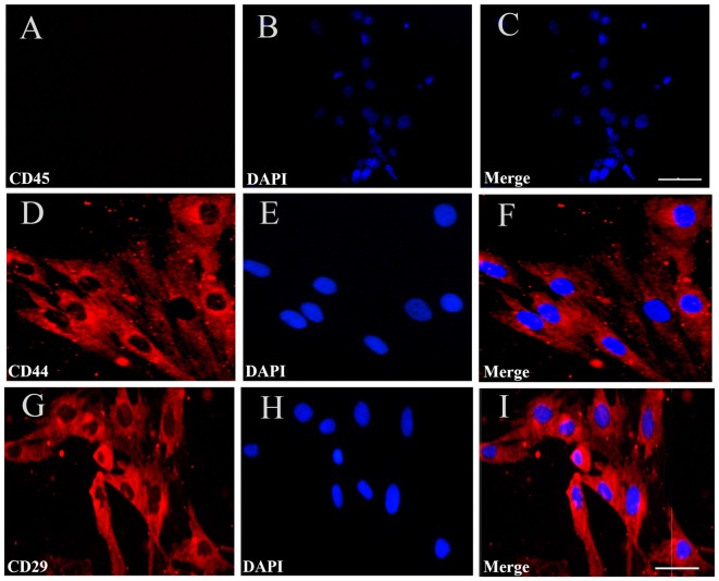
Figure 2.
Immunofluorescent staining for BMSCs identification. (A) Immunofluorescent staining of CD45 in the cultured BMSCs, but no cells expressed CD45. (D,G) There were CD44 and CD29 red positive immunofluorescence (IF). (B,E,H) Cell nucleus was stained by DAPI with blue fluorescence. (C,F,I) The merged images showed the mean positive rate of CD44, CD29and CD45 were about 98.8%, 99.3% and 0%. Bar = 50 μm in (A–C), 25 μm in (D–I). The mean positive rate represented as mean number of the positive cells/the number of DAPI was analyzed using Image-Pro plus 6.0 software. CD44, CD 29, CD45 are the specific surface markers to identify the BMSC.

