
FIGURE 4.
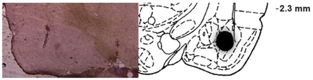
Infusion placements into the BLA. Representative photomicrograph illustrating placement of a cannula and needle tip, and schematic diagram of a coronal section of the rat brain (anteroposterior, –2.8 mm from bregma), adapted from the atlas of Paxinos and Watson (2007), depicting the diffusion of methylene blue in the BLA for rats included in the statistical analysis.