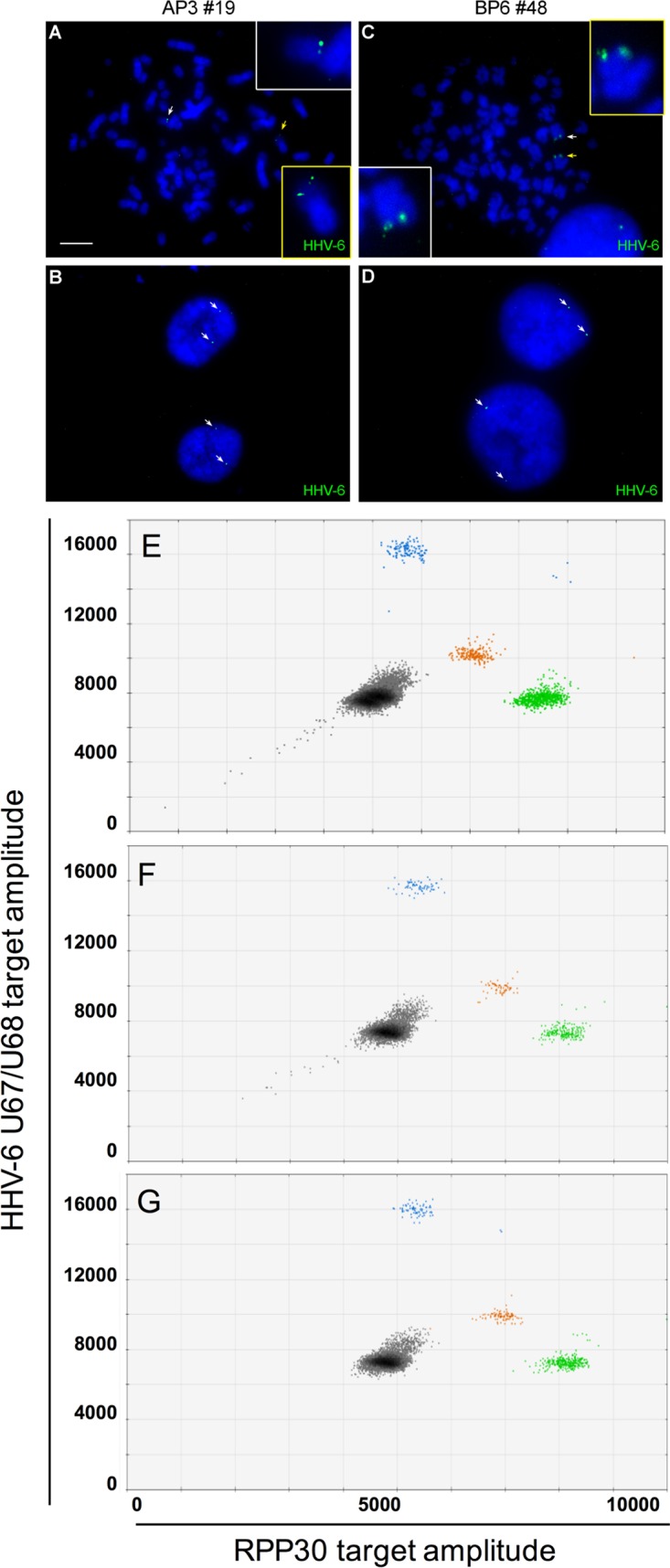
FIG 5.

Generation of HHV-6A and HHV-6B doubly integrated U2OS cells. U2OS-AP3 (ciHHV-6A+) and U2OS-BP6 (ciHHV-6B+) were superinfected with HHV-6B and HHV-6A, respectively, and submitted to single-cell cloning. Clones positive for two viruses were identified by FISH and ddPCR. Metaphase spreads (A and C) and interphase nuclei (B and D) of AP3#19 (A and B) and BP6#48 (C and D) showing the presence of two distinct HHV-6 hybridization signals (arrows). (E and G) Droplet digital PCR analysis of cell lines containing integrated HHV-6A and integrated HHV-6B. (E) ciHHV-6A+ and ciHHV-6B+ cell lines assayed together to produce a droplet plot with multiple clusters. Gray droplets are negative for template. Blue droplets are positive for HHV-6A. Orange droplets are positive for HHV-6B. Green droplets are positive for RPP30. (F) ddPCR analysis of the U2OS AP3 superinfected with HHV-6B to generate clone 19 containing both HHV-6 species. On average, this clone contained 2.9 HHV-6A and 2.2 HHV-6B genomes/cell. (G) ddPCR analysis of the U2OS BP6 clone superinfected with HHV-6A, resulting in clone 48 containing both HHV-6 species. On average, this clone contained 0.6 HHV-6A and 0.6 HHV-6B genomes/cell.
