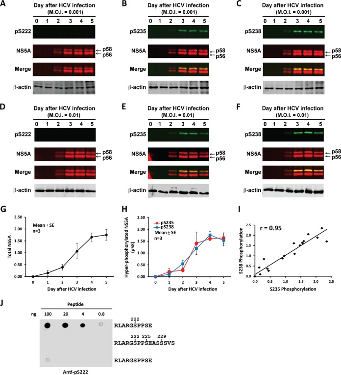
FIG 2.
NS5A S235 phosphorylation and S238 phosphorylation showed parallel time-dependent increases in the infected cells. (A to F) Immunoblots for NS5A phosphorylation at S222, S235, and S238 in the HCV-infected Huh7.5.1 cells at an MOI of 0.001 (A to C) or 0.01 (D to F). ß-Actin served as a loading control. (G and H) Line plots summarizing total NS5A and NS5A phosphorylation at S235 and S238 from three independent experiments. Relative protein abundance was quantified with the Li-Core Odyssey scanner and software. Values are means ± standard errors (SE). (I) Pearson's correlation analysis for S235 and S238 phosphorylation. (J) Dot blot analysis of the S222 phosphorylation-specific antibody for detection interference by phosphorylation at S225 and S229. Phosphorylated serine residues are numbered.

