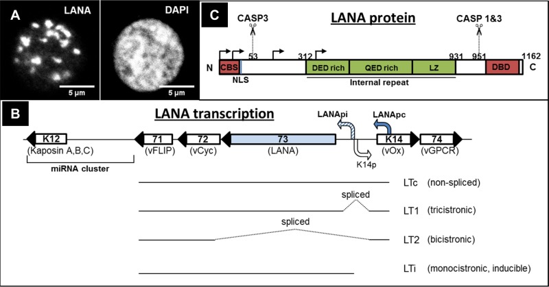
FIG 1.
(A) Immunofluorescence staining of LANA speckles in PEL cells. (Left panel) LANA staining. (Right panel) DAPI (4′,6-diamidino-2-phenylindole) staining of the nucleus. (B) Simplified structure of KSHV latency locus, indicating the LANA constitutive promoter (Lana pc) and the bidirectional lytic LANA inducible (LANA pi)/K14 promoter, as well as transcripts produced from the latency locus (20, 22, 26, 27). Viral open reading frame (ORF) numbers are indicated inside the respective genes, and the customary names are given below. ORF71 (vFLIP—viral FLICE inhibitory protein), ORF72 (vCyc—viral cyclin), ORF73 (LANA), ORF K14 (vOx—viral homologue of OX2), ORF74 (vGPCR—viral G protein coupled receptor) are indicated. (C) Schematic representation of LANA domains and important motifs. Caspase 1 and 3 cleavage sites (CASP1/3) (13) are also indicated. Arrows indicate canonical and alternative translation initiation sites (17). CBS, chromatin binding site; NLS, nuclear localization signal; DBD, DNA binding domain; LZ, leucine zipper.

