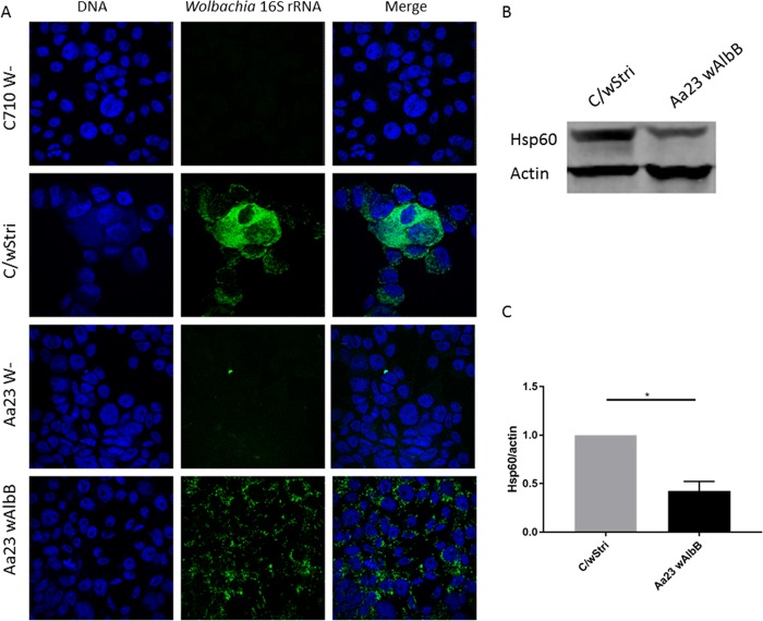
FIG 2.
Wolbachia wAlbB and wStri lines are infected at similar frequencies but different densities. (A) Fluorescent in situ hybridization of Aa23 cells with and without wAlbB and C710 cells with and without wStri. Using the differential interference contrast (DIC) channel to delineate cell barriers, cells were counted and recorded as Wolbachia infected or Wolbachia free to determine the frequency of Wolbachia infection in each cell line. Data are recorded in Table 1. (B) Western blot to quantitate Wolbachia density (Hsp60) relative to host (actin) proteins. (C) Hsp60 normalized to actin band intensity was quantified for three independent experiments to compare wStri density to wAlbB density in A. albopictus cells (significance, P < 0.05). wStri density is normalized to 1 for each experiment to compare wAlbB density. Statistical significance was determined by paired t test (one-tailed; alpha = 0.05) on the natural log of the (Hsp60/actin) ratio accounting for nonnormal distribution of fluorescent intensities. Statistical tests were calculated by GraphPad Prism.

