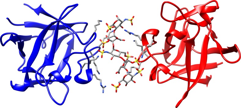
Fig. 1.
Detail of the crystallographic complex FGF1-HE dp5-FGF1 (PDB ID 1AMX, 3.0 Å) showing each protein chain (FGFA and FGFB) in red and blue cartoon and the HE dp5 ligand in sticks and coloured by atom. The side chains of the protein positively-charged residues in the binding interface are displayed in grey sticks

