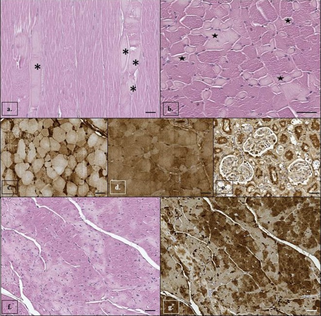
Fig. 3.
Stenella coeruleoalba. Skeletal muscle (longissimus dorsi): myocytes have (a, b) acute degenerative changes characterized by fibers hyaline degeneration (asterisks) and multifocal loss of cross striations and mild endomisial edema (stars) probably due to an acute ischemic damage. Hematoxylin and eosin staining. Scale bar=50 µm. Immunohistochemical analysis of skeletal muscle (longissimus dorsi): (c) strong intracytoplasmatic positive immunoreaction for fibrinogen in affected myocytes and (d) depletion of myoglobin in degenerated myocytes is demonstrated by decreased immunoreaction for myoglobin. Mayer’s hematoxylin counterstain. In kidney (e), the apical cytoplasm of degenerated tubular cells and granules in the Bowman space and tubular lumen show positive immunoreaction for myoglobin. Mayer’s hematoxylin counterstain. Scale bar=50 µm. Cardiac muscle: (f) cardiomyocytes present multifocal hyaline degeneration and loss of cross striations. Hematoxylin and eosin staining. Scale bar=50 µm. Immunohistochemical analysis of cardiac muscle: (g) decreased immunoreaction for myoglobin in degenerated cardiomyocytes. Mayer’s hematoxylin counterstain. Scale bar=50 µm.

