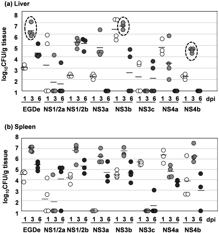
Fig. 2.
Bacterial growth of 7 serotypes of L. monocytogenes. (a) Bacterial growth in the liver. (b) Bacterial growth in the spleen. Mice were inoculated intravenously with each strain. At 1, 3 and 6 dpi, the livers and spleens were harvested from 3 or 4 mice for each strain, and live bacteria were counted. The detection limit was 102 CFU/g tissue. The bacterial counts in the livers of EGDe-, NS3b- and NS4b-infected mice at 3 dpi were enclosed by dashed circles for emphasis. White dots, 1 dpi; light gray dots, 3 dpi; dark gray dots, 6 dpi. Bars indicate the means of three or four samples.

