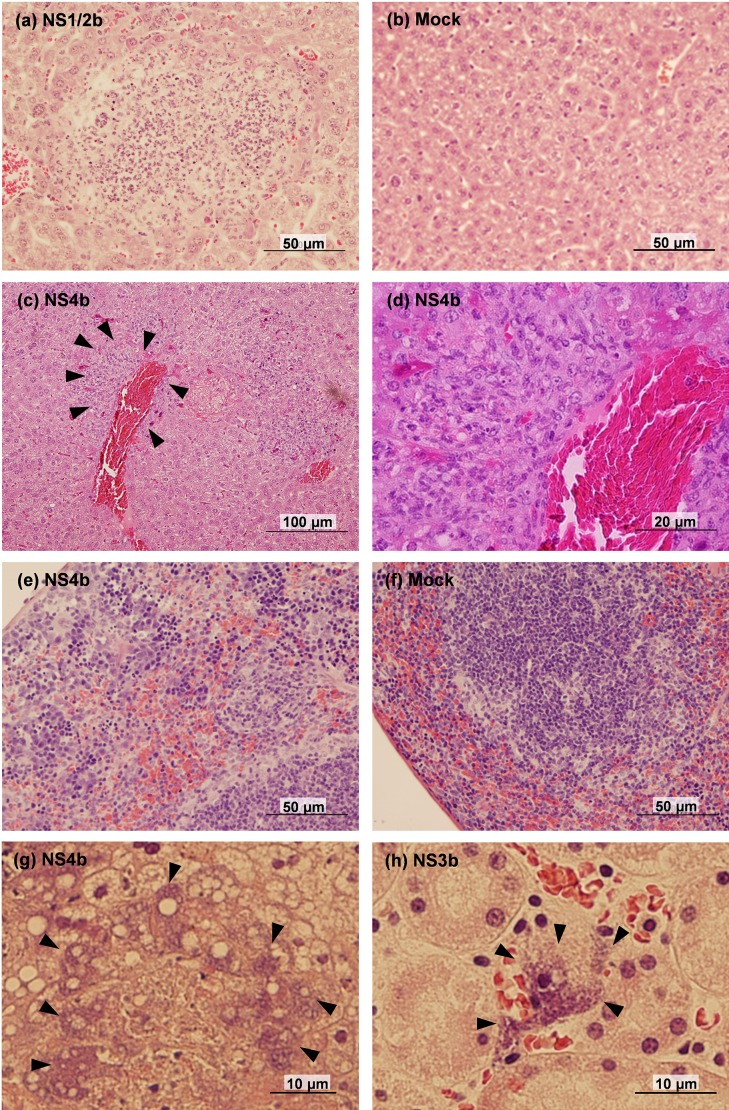
Fig. 3.
Histopathology of L. monocytogenes-infected Balb/c mice. (a) Abscess in the liver of NS1/2b-infected mouse. (b) Liver of mock-infected mouse. (c) Perivascular infiltration of macrophages in the liver of NS4b-infected mouse. Arrowheads surround the area with macrophage infiltration. (d) Magnified image of the area surrounded by arrowheads in (c). (e) Migration of macrophages in the splenic sinus of NS4b-infected mouse. (f) Spleen of mock-infected mouse. (g, h) Bacterial clumps in the dead mice, observed in the liver of NS4b-infected mouse (g) and in the kidney of NS3b-infected mouse (h). Arrowheads show the bacterial clumps.

