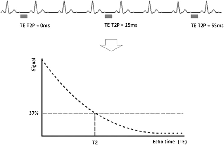
Fig. 2.
Principles of T2 mapping with different T2 preparatory durations with a long repetition time between the used cardiac cycles, crucial to allow as complete T1 recovery as possible, followed by reconstruction of the transverse relaxation curve in each pixel assuming satisfactory registration. T2 is defined as the time in milliseconds by which the transverse magnetisation has decayed to 37% of the original value. Many distorting factors are not illustrated, and many T2 mapping sequences ‘fill the gaps’ with gradient activity without RF, so that the patient does not consider the scan complete and start breathing too early

