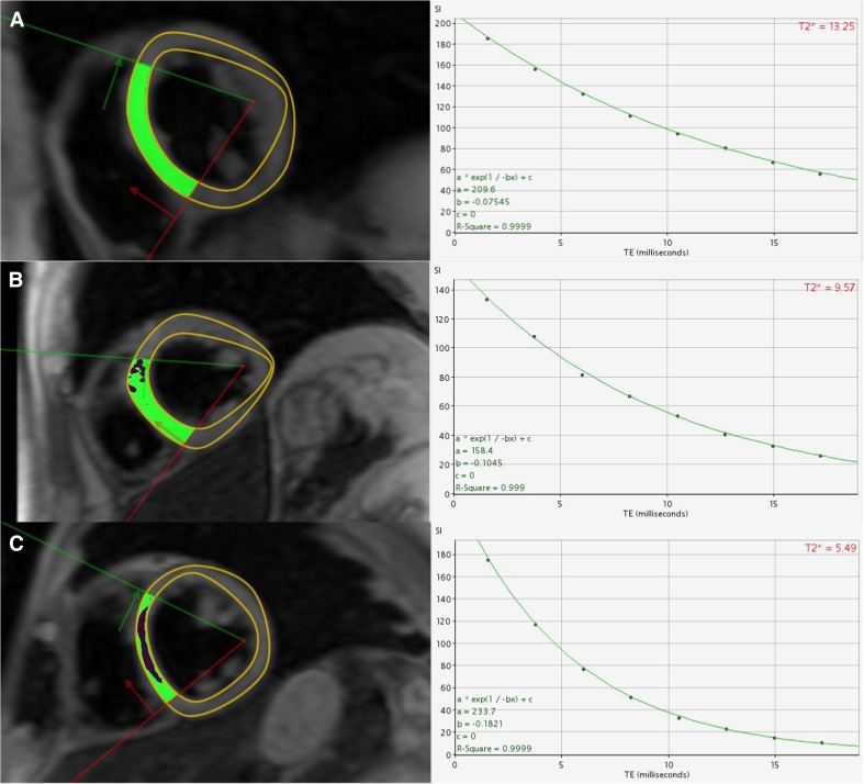
Fig. 3.
T2* transverse relaxation curves in three separate patients with mild >14 ms (a), moderate 10–14 ms (b) and severe <6 ms (c) iron overload. Black blood T2* imaging is used rather than white blood due to superior reproducibility and reduced imaging artefact [61]

