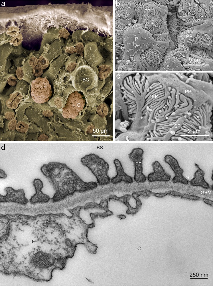
Fig. 1.
Glomerular ultrastructure. a False-colored low-resolution scanning electron micrograph of mouse renal cortex displaying several tubuli (T) and two glomeruli (G). In addition, an empty Bowman’s capsule (BC) can be seen from which the glomerulum was lost during preparation. b Detailed scanning electron micrograph showing a podocyte (P), primary processes (PP) originating from the cell body and foot processes (FP). c High-resolution scanning electron micrograph illustrating primary processes (PP), secondary processes (SP) and foot processes (FP). d Transmission electron micrscopy of a mouse glomerular capillary (C) revealing the structural composition of the glomerulus. The endothelial cell (E) coats the inner surface of the capillary wall and is followed by the three layers of the glomerular basement membrane (GBM). On the outside of the capillary, the foot processes (FP) cover a major part of the GBM circumference. The slit-diaphram can be discerned in between the foot processs. The primary filtrate drains into Bowman’s space (BS) and, via the urinary pole, reaches the proximal tubule

