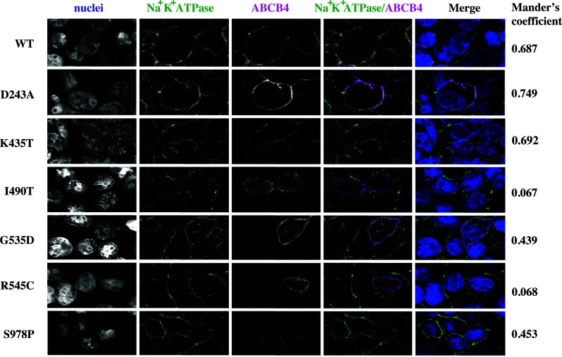
Fig. 2.

Confocal analysis of ABCB4 variant sub-cellular localisation. Trafficking of ABCB4 mutants D243A, K435T, G535D, R545C, I490T, and S978P was examined by confocal microscopy (shown in magenta in the merged images) in cells co-expressing ATP8B1/CDC50. The Na+/K+ ATPase, shown in green in the merged images, marks the plasma membrane. Co-localisation in the merged image is white when the pixels for the Na+/K+ ATPase and ABCB4 overlap and have the same intensity. The Mander’s coefficient for the given field of view provides a measure of this co-localisation above a calculated threshold, irrespective of pixel intensity
