
Fig. 4.
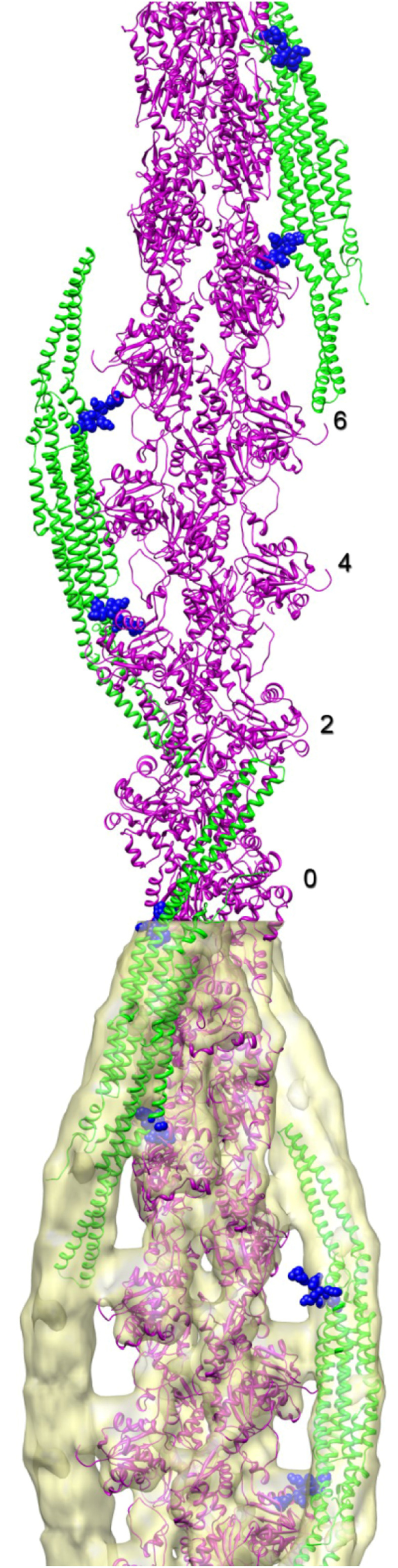
Model for pacsin-2 bound to F-actin, based on EM reconstruction of F-actin decorated by pacin-2 [50]. Actin subunits (magenta ribbons) are numbered along one strand. The two green pacsin-2 ribbons on the right bind to that strand. The green pacsin-2 ribbon on the left binds to the opposite actin strand. The yellow surface at the bottom is a three-dimensional reconstruction of the atomic model shown, after imposing the actin helical symmetry and filtering to 12 Å resolution. Residues of the wedge loop, pointing towards F-actin, are represented as blue spheres