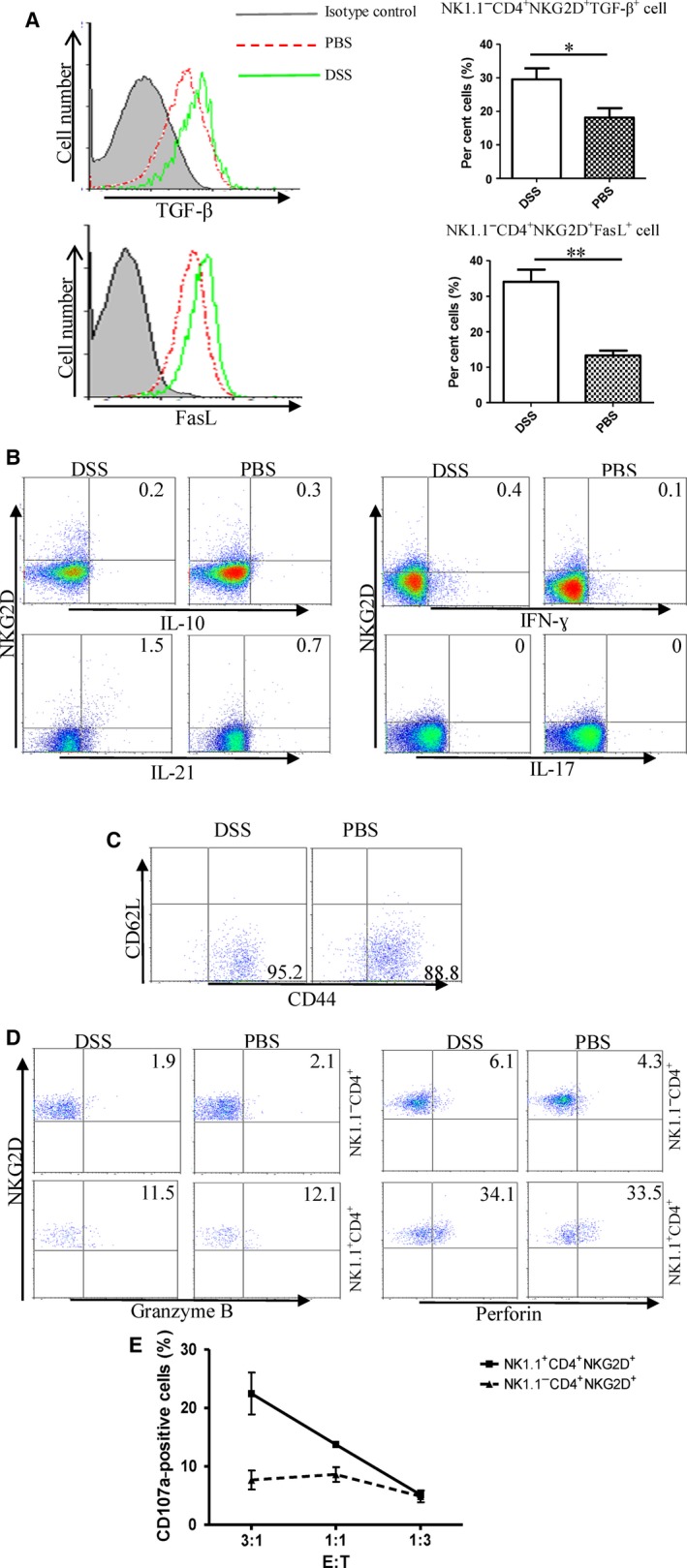
Figure 2.

Capacity of cytokine production and cytotoxicity of NK1.1− CD4+ NKG2D+ cells. (A) NK1.1− CD4+ NKG2D+ cells were stained by TGF‐β and FasL antibody. (B) NK1.1− CD4+ NKG2D+ cells were intracellularly stained by IL‐10, IFN‐γ, IL‐21 and IL‐17 antibodies after stimulation by PMA and ionomycin. (C) Costaining of CD62L and CD44 on NK1.1− CD4+ NKG2D+ cells. (D) Intracellular staining of granzyme B and perforin. (E) Degranulation of NK1.1− CD4+ NKG2D+ and NK1.1+ CD4+ NKG2D+ cell after incubating with B16‐MICA target cells is measured by CD107a expression on cell membrane. All experiments were performed three times.
