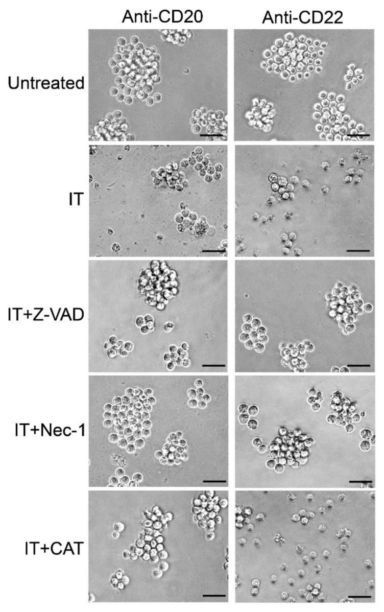
Figure 7.
Morphological analysis of Raji cells assessed using phase-contrast microscopy. Cells were treated for 96 h with 1 nM anti-CD20 IT (left) or 0.01 nM anti-CD22 IT (right) alone (IT) or in the presence of 10 μM pan-caspase inhibitor (Z-VAD), 10 μM necroptosis inhibitor necrostatin-1 (Nec-1), or 10 U/mL hydrogen peroxide scavenger catalase (CAT). Z-VAD, Nec-1 and CAT were added 3 h before the ITs. Untreated cultures were grown in the absence of ITs. Magnification, 400 ×. Scale bars correspond to 50 µm.

