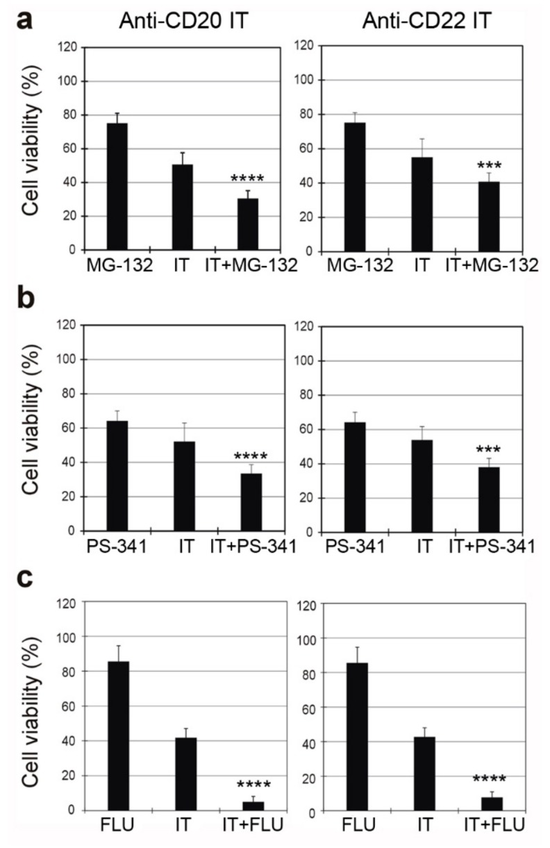
Figure 8.
Viability of Raji cells treated for 96 h with 1 nM anti-CD20 IT (left) or 0.01 nM anti-CD22 IT (right) alone or in the presence of the proteasome inhibitors MG-132 (0.1 μM) (a) or PS-341 (1 nM) (b) or the purine analogue fludarabine (FLU) (0.75 μM) (c). Inhibitors and FLU were added 3 h before the ITs and maintained for the entire incubation time. Cell viability was determined after 96 h. Means ± S.D. of three independent experiments, each in triplicate, are showed as the percentage of the untreated cell values. Statistical significance was determined by ANOVA/Bonferroni test. Asterisks indicate the significant difference in each experimental condition between IT alone and IT plus the inhibitor or the purine analogue (**** p < 0.0001; *** p < 0.001).

