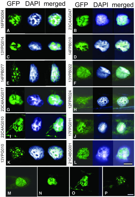
Figure 3.
Localization of the GFP-Fused Proteins of the Unknown Protein Group in Allium Epidermal Cells.
The images of the GFP fusion proteins are shown at the left, the nuclei stained with DAPI are in the middle, and the merged images with these two are at the right. The GFP figures represent localization of several unknown proteins fused to GFP. For detailed characteristics of these proteins, see Table 3. Bar = 25 μm.
(A), (C), (E), (G), (I), and (K) Proteins localized in an inner nuclear matrix region.
(B), (D), (F), (H), (J), and (L) Proteins colocalized with DNA/chromatin.
(M) to (P) Figures reduced to show whole cytoplasmic area of the cells in (F), (G), (I), and (K), respectively.

