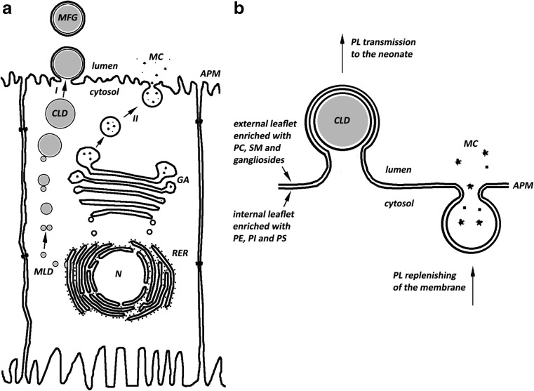
Fig. 3.
Scheme of secretion of milk components (Fig. 3a – based on Ref. [72]). In Fig. 3b, arrows indicate the flux of phospholipids to and from the apical membrane of the mammary gland. In this model milk fat globules and water soluble milk components are secreted separately, by two distinct pathways (I and II, respectively). These two pathways enable replenishing the phospholipid content of the apical membrane, maintaining its constant composition and, thereby, the continuous secretion of milk components. Abbreviations: MFG-milk fat globule, CLD-cytoplasmic lipid droplet, MLD-microlipid droplet, N-nucleus, MC-milk components, APM-apical plasma membrane, GA-Golgi apparatus, RER-rough endoplasmic reticulum, PL-phospholipid

