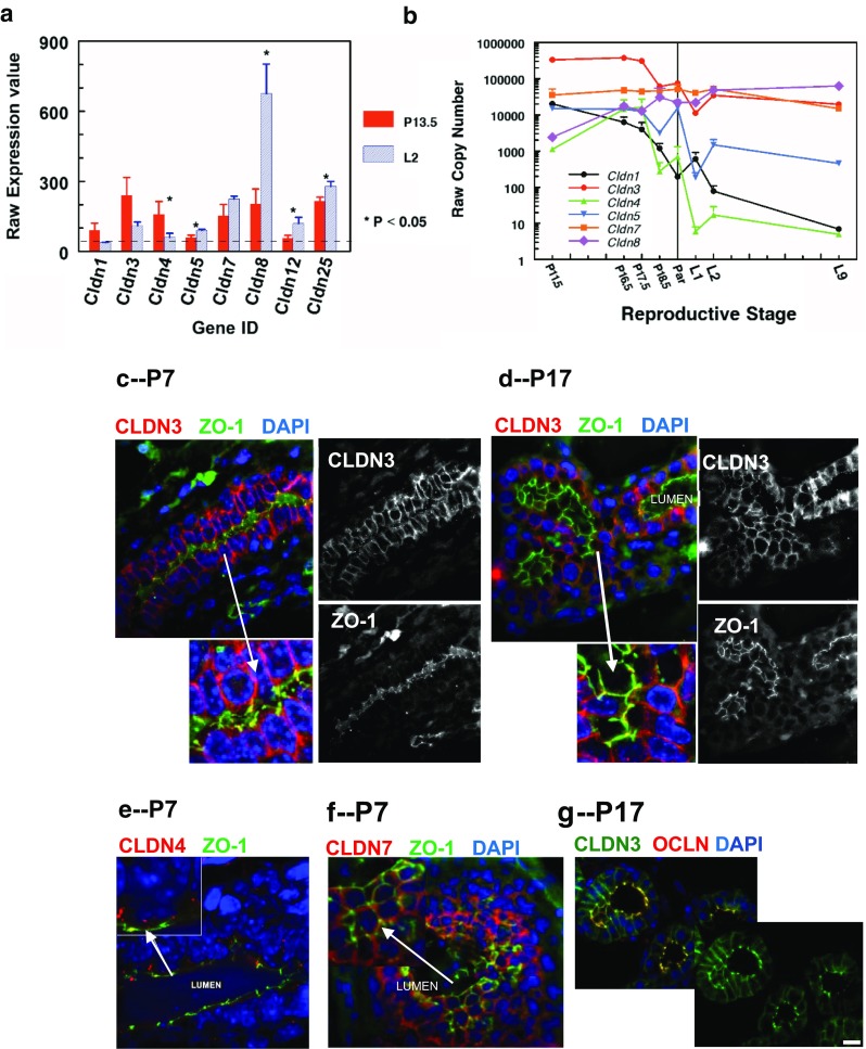
Fig. 4.

Expression and localization of claudins in pregnancy. a. Microarray analysis of claudin mRNA in MECs isolated from pregnant (day 13.5) and lactating (day 2) FVB mice [58]. MECs were isolated from the 4th mammary gland according to the techniques of Rudolph and colleagues [58]. mRNA expression was analyzed using Affymetrix MoGene_1_0-st-v1 chip arrays [77]. Raw expression values for all claudins with expression values above 40 (dotted line) at pregnancy day 13.5 (red) or lactation day 2 (blue) are shown. * Expression significantly different between pregnancy and lactation, P < 0.05 b. mRNA expression of claudins 1, 3, 4, 5, 7 and 8 during the transition from pregnancy to lactation by quantitative real time PCR using lysates of whole mammary glands (See Methods, Fig. 4). d,e. Immunofluorescence localization of claudins-3, −4 and −7 in mammary glands from CD1 pregnant mice (Neville laboratory, see Methods section). c,e,f. Pregnancy day 7. Similar results were observed in both formalin-fixed (shown) and frozen (not shown) sections. d,g. Pregnancy day 17. g. Localization of claudin-3 in the mammary gland from the ICR mouse; in this strain claudin-3 was localized both basolaterally and with occludin at pregnancy day 17 [43]. c-g. Scale bars, 20 μm
