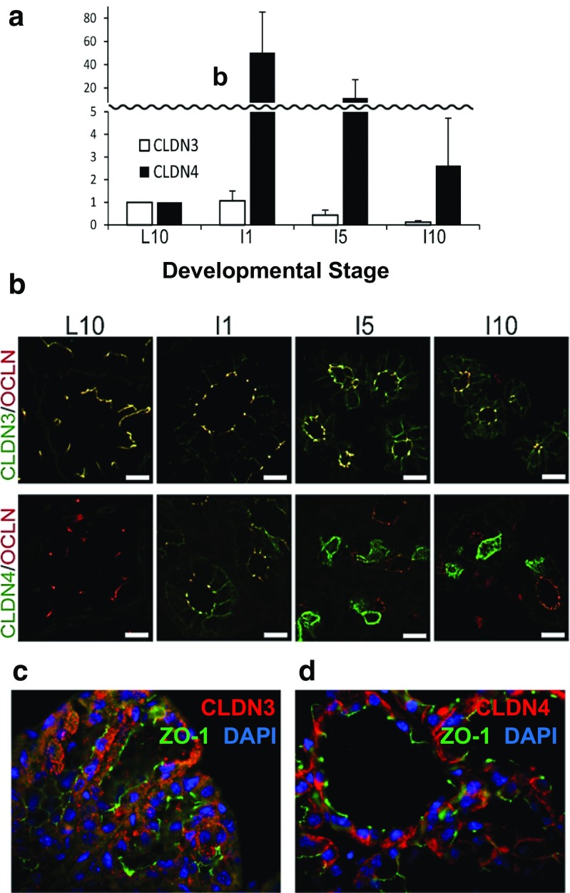
Fig. 7.
Claudins in mammary involution. a Levels of claudins-3 and -4 protein from Western blots during involution. Pups were removed from day 10 lactating ICR mice. Dams were sacrificed at day 10 of lactation and 1, 5 and 10 days later. Proteins from minced mammary glands were electrophoresed and visualized with appropriate antibodies (ThermoFisher) as described [43]. b Immunofluorescence of claudins-3 and -4 in the samples from panel a compared to localization of the tight junction protein, occludin (OCLN). Images from Kobayashi lab after ref. [43]. c,d Higher power images from sections of mammary glands from 10 day lactating FVB mice sacrificed 2 days after pup removal and stained using antibodies for claudin-3, claudin-4 (claudins in red) and the tight junction protein ZO-1 (green). Nuclei stained with DAPI (blue) Significant cytoplasmic stain can be observed for both claudins with little or no overlap with ZO-1. See methods for preparation of these images from the Neville laboratory

