Fig. 8.
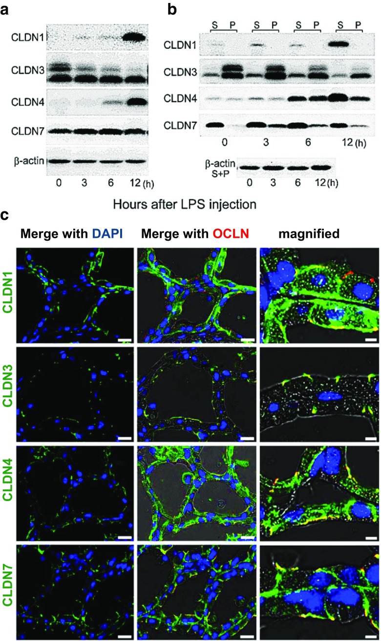
Effect of LPS on amount, type and localization of claudin protein. a. Western blot of claudins-1, −3, −4, and −7 in 10 day lactating gland 0, 3, 6, and 12 h after injection of E. coli lipopolysaccharide (LPS) into the teat canal of the fourth mammary gland of ICR mice. b. Western blot of claudins in Triton-X soluble (S) and insoluble (P) fractions of mammary gland lysates after LPS injection. c. Immunofluorescence analysis of claudins in the mammary gland 12 h after injection of LPS. Figures from ref. [31] Plos One

