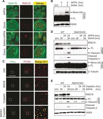
Figure 4.

A) Colocalization of labeled DNA‐Pt with RAD18 in U2OS cells. Cells were treated with APPA (250 μm for 3 h) and SAHA (2.5 μm for 5 h). Zoomed images are 3×. Scale bar, 20 μm. B) Western blot analysis of PCNA mono‐ubiquitination in U2OS cells. Cells were treated with APPA (250 μm) and SAHA (2.5 μm) as indicated. C) Focal accumulation of Polη colocalizing with PCNA in U2OS cells. Cells were treated with cisplatin (10 μm for 3 h) and SAHA (2.5 μm for 5 h). Zoomed images are 6×. Scale bar, 20 μm. D) and E) Western blot analysis of apoptotic markers in WT and RAD18 KO HCT‐116 cells. Cells were treated with APPA (250 μm), SAHA (2.5 μm) and cisplatin (10 μm) as indicated. FL, full length.
