
Figure 2.
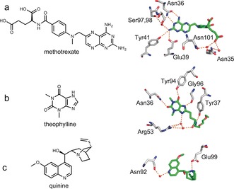
Antibody‐binding of methotrexate (a), theophylline (b), and quinine (c). The chemical structures of the three drugs are shown on the left. The crystal structures of antibodies with the methotrexate (PDB ID: 4OCX), theophylline derivative (PDB ID: 5BMF), and quinine (PDB ID: 4UIN) were superimposed, showing the interactions of the residues with the ligands (carbon atoms in green). Extensive hydrogen bonding (dotted orange lines) links the three antigens to the antibodies, either directly or via water molecules (red balls).