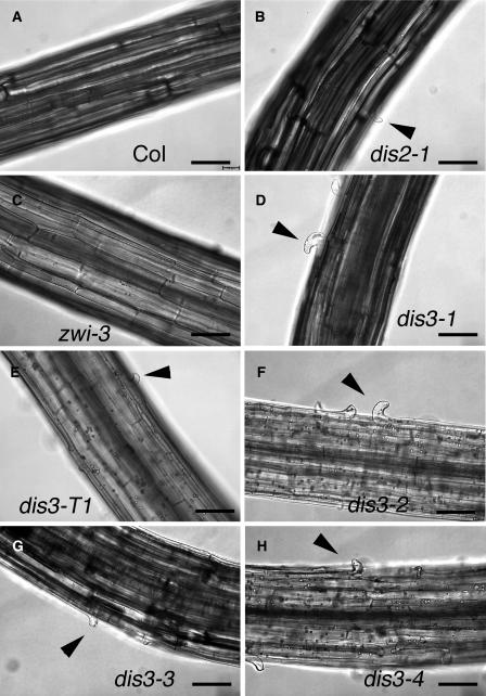
Figure 2.
Cell–Cell Adhesion Defects in Etiolated Hypocotyls of dis3 Alleles.
(A) Wild-type hypocotyls with adherent epidermal cells.
(B) Epidermal cell–cell adhesion defects in dis2-1 (arpc2).
(C) zwi-3 hypocotyls with adherent epidermal cells.
(D) Epidermal cell–cell adhesion defects in dis3-1.
(E) Epidermal cell–cell adhesion defects in dis3-T1.
(F) Epidermal cell–cell adhesion defects in dis3-2.
(G) Epidermal cell–cell adhesion defects in dis3-3.
(H) Epidermal cell–cell adhesion defects in dis3-4.
Differential interference contrast images of whole-mounted, 7-DAG, dark-grown seedlings. Arrowheads indicate the location of cell–cell adhesion defects. Bar = 100 μm in all panels.

