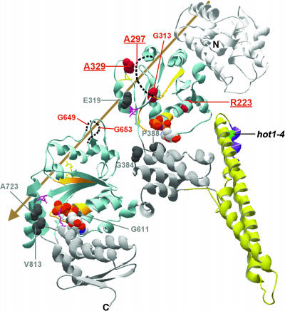
Figure 9.
Location of the hot1-4 Suppressor Mutations on the T. thermophilus ClpB Monomer Structure.
The major domains and motifs of HCP100/ClpB are colored as indicated and described in Figure 8. The sites of Class 1 suppressor mutations are space-filled dark gray, and sites of the Class 2 suppressor mutations are space-filled in red. The three strongest Class 2 suppressors are indicated in bold and are underlined. For A297, G653, and G649, which are in unresolved segments of the structure, dotted lines are used to indicate the relative positions of those segments. The arrow indicates the general position of the axial channel of the hexameric form of the protein. Residue numbering corresponds to AtHsp101. Figure was prepared with Swiss PDB viewer.

