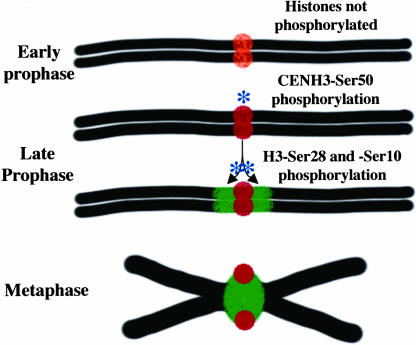
Figure 8.
A Kinase Diffusion Model for Pericentromere Determination.
At top is a prediplotene chromosome and its centromere with unphosphorylated CENH3 (orange). At diplotene, a histone kinase phosphorylates CENH3 first (red), then travels outward over the pericentromere and phosphorylates histone H3 (green) in a diffusion-limited manner. The phosphorylated CENH3 interacts with the spindle, whereas phosphorylated histone H3 marks the pericentromere and serves to enhance or stabilize cohesion deposition.

