
Figure 5.
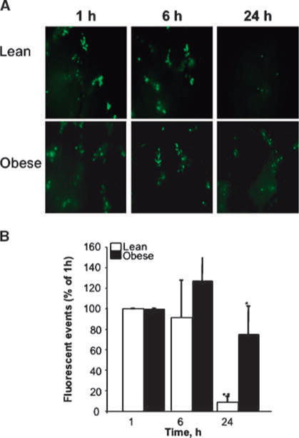
Persistent detection of tumor cells in livers of obese mice. MC-38 cells were labeled with a fluorescent dye (CMFDA), 3 × 10⁵ cells were injected per mouse through the intrasplenic/portal route, and mice were splenectomized 1 min later. Livers were dissected at the indicated time points after tumor cell injection and imaged by inverted fluorescent videomicroscopy (Leica DM IRB) at ×100 magnification (A). Ten random frames were captured and analyzed using OpenLab software to define and count fluorescent (green) CMFDA tumor events larger than 10 pixels. Columns, mean total number of events per liver per mouse (n = 5 mice per group) per time point; bars, SEM. *, P < 0.05 as compared to lean animals at the same time point; **, P < 0.05 as compared to lean animals at 1 h after cell injection.