
FIGURE 4.
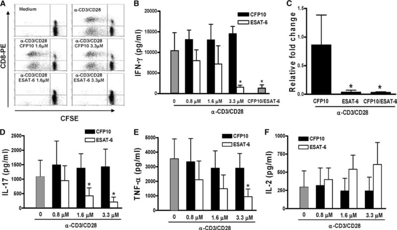
Effect of ESAT-6 on proliferation and cytokine production by CD3+ cells stimulated with anti-CD3 and anti-CD28. A, Purified CD3+ cells were labeled with CFSE and cultured in medium alone (top right panel), CFP10 (middle panels), or ESAT-6 (lower panels) for 1 h, before addition of anti-CD3 (10 μg/ml) and anti-CD28 (1 μg/ml). The top left panel shows cells cultured in medium alone, without anti-CD3 or anti-CD28. After 72 h, the cells were stained with anti-CD8-PE, and CFSE content was analyzed by flow cytometry. One representative result of experiments performed for three donors is shown. B, Purified CD3+ cells from five healthy donors were cultured with medium alone, ESAT-6, CFP10, or ESAT-6/CFP10 heterodimer (3.3 μM each, far right bar) for 1 h, before addition of anti-CD3 and anti-CD28, as in A. After 48 h, IFN-γ levels in the culture supernatants were measured by ELISA. Mean values and SD are shown. *, p < 0.001, compared with the cells treated with anti-CD3 plus anti-CD28 mAbs and equimolar concentration of CFP10. C, Purified CD3+ cells from three healthy donors were cultured as described in B for 1 h, before addition of anti-CD3 and anti-CD28. After 48 h, IFN-γ mRNA expression was measured by real-time PCR. *, p < 0.005, compared with the cells stimulated with anti-CD3 plus anti-CD28 in the presence of CFP10. D–F, Production of IL-17 (D), TNF-α (E) and IL-2 (F) by purified CD3+ cells, cultured as described in B. Mean values and SD from five subjects (IL-17), and eight individuals (TNF-α and IL-2), respectively, are shown. *, p < 0.005 (both D and E), compared with cells stimulated with anti-CD3 plus anti-CD28 in the presence of CFP10.