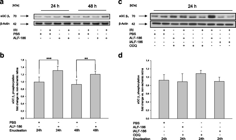
Fig. 2.

Effect of ALF-186 treatment on the expression of soluble guanylyl cyclase (sGC) β1. a Representative western blot image (n = 8) showing the increase of sGC β1 compared to β-Actin after immediate ALF-186 postconditioning. Enucleation was performed either 24 or 48 h after IRI. b Densitometric analysis of n = 8 western blots for sGC β1 after ALF-186 (data are mean ± SD; IRI vs. IRI + ALF-186 24 h, *** = p < 0.001 and IRI vs. IRI + ALF-186 48 h, ** = p < 0.01). c Representative western blot image and densitometric analysis of n = 8 Western Blots for sGC β1 compared to β-Actin after ODQ inhibition and ALF-186, ODQ inhibition alone and ODQ-inhibition and iALF-186. Enucleation was performed 24 h after IRI. d Densitometric analysis of n = 8 Western Blots for sGC β1 after ALF-186 in the presence of ODQ
