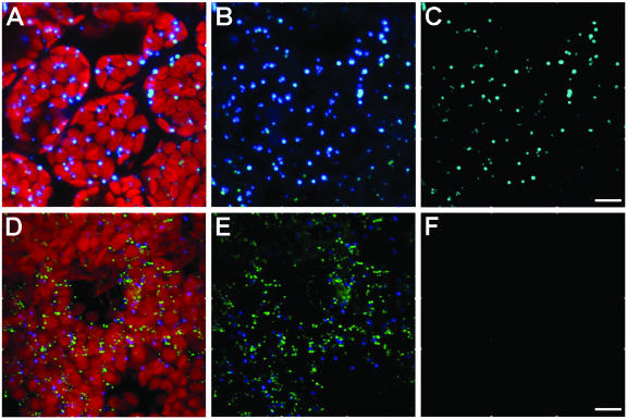
Figure 5.
Immunofluorescence colocalization evaluations of catalase and HMGR in cotyledons of parenchyma cells of Arabidopsis seedlings. A to C, Colocalization analysis of confocal projections of mouse anti-tobacco catalase and rabbit anti-cottonseed catalase. Punctate mouse anti-catalase and rabbit anti-catalase shown with (A) and without (B) autofluorescent red plastids, and the binary mask result showing the colocalization of both antibodies in punctate peroxisomes (C). D to F, Colocalization evaluations of confocal projections of mouse anti-tobacco catalase (blue) and rabbit anti-CD1 (green). Triple-fluorescence images of both antibodies and plastids of a group of parenchyma cells (D), the combination of the antibody images only (E), and the binary mask colocalization analysis showing the absence of colocalizing pixels (F). Images are three-dimensional projections of a series of 40 to 50 sections with a z step of 0.25 μm. Bars = 10 μm.

