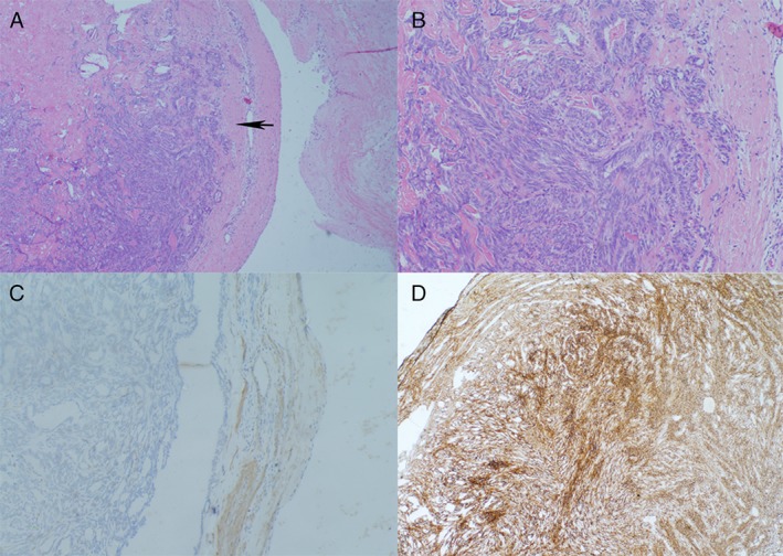
Figure 3.

Pathology of the tumour. (A) Haematoxylin and eosin (HE) stain of the tumour shows that the tumour arose from the intimal of pulmonary artery (50×). The black arrow indicates the neoplasm from the artery wall. (B) Microscopically, the tumour was composed of pleomorphic spindle cells (HE stain, 200×). (C) Tumour cells were immunohistochemically positive for α‐smooth muscle actin (SMA). (D) Immunostaining for vimentin (VIM) highlights numerous neoplasm cells derived from mesenchymal tissue.
