. 2017 May 29;40(2):398–407. doi: 10.1590/1678-4685-GMB-2016-0213

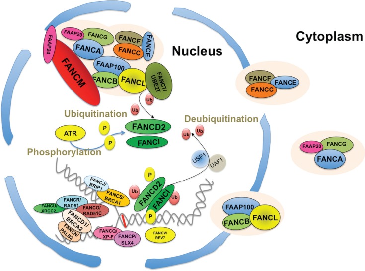
Figure 1. Schematic representation of the subcellular distribution of the FANC proteins, their association and their relocalization in nuclear foci at stalled replication forks. In unstressed conditions, three subcomplexes are present in the nucleus and/or the cytosol: FANCA, FANCG and FAAP20; FANCC, FANCE and FANCF; and FANCB, FANCL and FAAP100. In the presence of DNA damage (the red line represents an interstrand crosslink) that leads to stalled replication forks, all the FANC proteins shuttle into the nucleus to form the FANCcore complex to monoubiquitinate FANCD2 and FANCI, which in turn assemble to subnuclear foci, where they colocalize with several other proteins involved in homologous recombination, including other FANC and FANC-like representatives. The USP1:UAF1 dimer deubiquitinates both FANCD2 and FANCI.

Figure 1