Fig. 1.

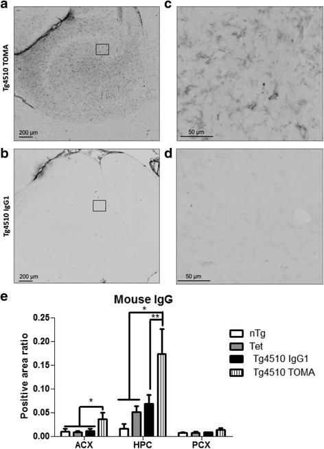
Tg4510 mice injected systemically with TOMA present elevated levels of mouse IgG in the brain. Micrographic representation of mouse IgG staining in hippocampus (HPC) of Tg4510 mice treated with TOMA (Tg4510 TOMA, a, c) or IgG1 (Tg4510 IgG1, b, d) for 4.5 months. c, d Magnification of square area in a and b, respectively. No staining was observed in nontransgenic or Tet-only littermates. Immunostaining quantification (e) utilizing Mirax software (Zeiss Inc.) in the anterior cortex (ACX), HPC, and posterior cortex (PCX) of nontransgenic littermates (nTg, white bars), tet-only mice (Tet, grey bars), and Tg4510 mice treated with IgG1 (Tg4510 IgG1, black bars) or TOMA (Tg4510 TOMA, striped bars) for 5 months. Two-way ANOVA showed a significant increase in positive area ratio stained for mouse IgG in the ACX and HPC of Tg4510 mice treated with TOMA compared to Tg4510 mice treated with IgG1 and nontransgenic and Tet-only control mice. Data presented as mean ± SEM, n = 10/group. *p < 0.05, **p < 0.01. Scale bar = 200 μm. TOMA tau oligomer monoclonal antibody
