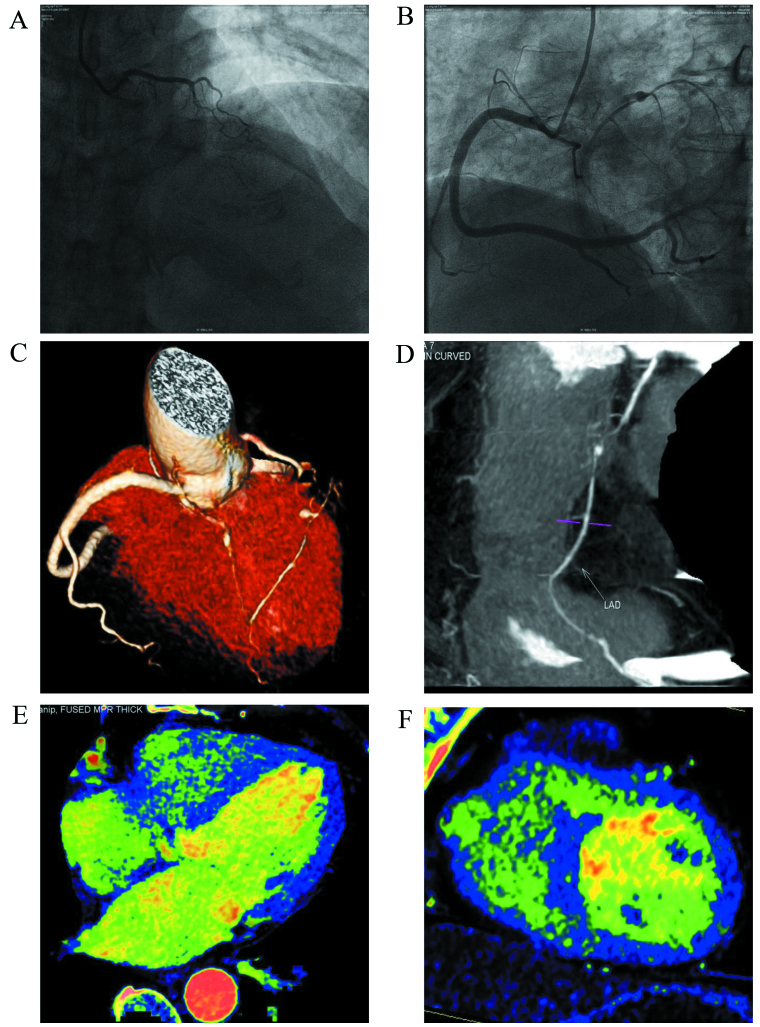
Figure 1.
A 63-year-old female complained of chest stuffiness. (A and B) Images indicate failure to visualize the middle segment of the anterior descending branch with coronary artery angiography upon admission (yellow arrow). The case was treated for an acute myocardial infarction due to an anterior descending branch occlusion. (C and D) Images indicate the small distal segment of the left anterior descending artery by computed tomography angiography plus dual-energy computed tomography (DECT) (yellow arrow). The blood was supplied to the anterior descending branch by the collateral circulation of the right coronary artery. An aneurysm was observed in the anterior descending branch (blue arrow). (E and F) Show myocardial iodine maps from DECT, indicating normal myocardial perfusion.

