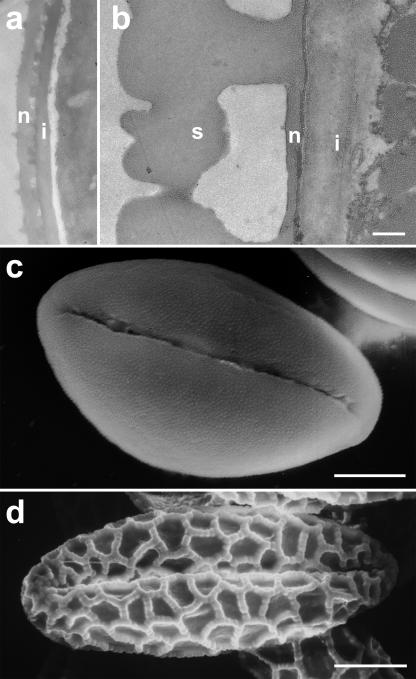
Figure 3.
Electron micrographs of the pollen grain cell wall structure. Transmission electron micrographs reveal that Solanum pollen grains (A) have a considerably thinner intine (i) than Lilium grains (B). While nexine (n) thickness is comparable, the sexine (s) of Lilium is extremely thick. Scanning electron micrographs show also that the Lilium sexine is structured in a coarse reticulate pattern (D), whereas Solanum is ornamented by very small scabrate structures (C). Bars = 0.5 μm (A and B); 5 μm (C); 20 μm (D).

