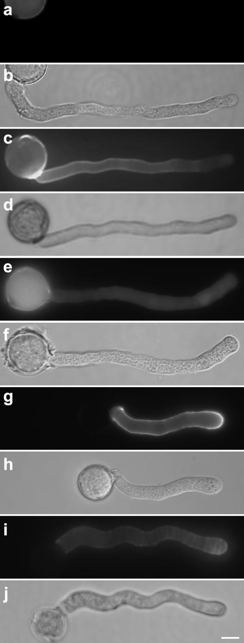
Figure 7.
Callose and pectin content in pollen tubes. A–F, Solanum pollen tubes labeled for callose with decolorized aniline blue and corresponding DIC images. Tubes grown in solidified medium (A and B) showed reduced abundance of callose compared to the control tubes grown in liquid medium (C and D). The presence of 1 mg mL−1 lyticase in liquid medium reduced the abundance of callose in the pollen tube (E) and caused an increase of the pollen tube diameter (F) compared to the control tubes (C and D). G to J, Solanum pollen tubes labeled for methyl-esterified pectins with monoclonal antibody JIM7 and corresponding DIC images. Pollen tubes grown in the presence of 1 mg mL−1 lyticase (I and J) showed significantly weaker label than the control cells (G and H). The distribution pattern remained the same, however, with a higher concentration of methyl-esterified pectin at the pollen tube apex. Bar = 10 μm.

