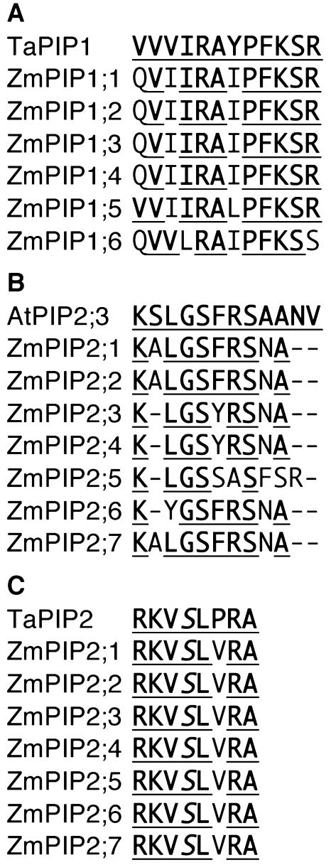
Figure 4.
A and B, Multiple alignment of the carboxy-terminal region of the Zm PIP1 and Zm PIP2 proteins and the TaPIP1 and AtPIP2;3 proteins, respectively. The consensus amino acids are underlined. C, Alignment of the conserved phosphorylation site of the PIP2 proteins. The TaPIP1, AtPIP2;3, and TaPIP2 sequences shown correspond to the sequence of the peptide used to make the respective antibodies.

