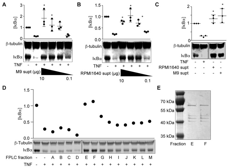
Figure 1.
ESF fractionation and identification. (A) IκBα immunoreactivity after incubating HCT-8 cells with ETEC H10407-M9 supernatant (0.1–10 µg protein) and then stimulating the cells with TNF (20 ng/mL, 20 min) Asterisks indicate significantly different IκBα abundance as compared with the ‘TNF only’ lane; (B) IκBα immunoreactivity after incubating HCT-8 cells with ETEC H10407-M9 supernatant (0.1–10 µg protein) and then stimulating the cells with TNF (20 ng/mL, 20 min); (C) IκBα immunoreactivity after incubating HCT-8 cells with ETEC H10407-RPMI1640 and ETEC H10407-M9 supernatants (10 µg protein) without TNF; (D) IκBα immunoreactivity after incubating HCT-8 cells with ETEC H10407-M9 supernatant FPLC fractions for 1.5 h and then stimulating the cells with TNF (20 ng/mL, 20 min); (E) Sliver staining of FPLC fractions E and F on 10% SDS-PAGE.

