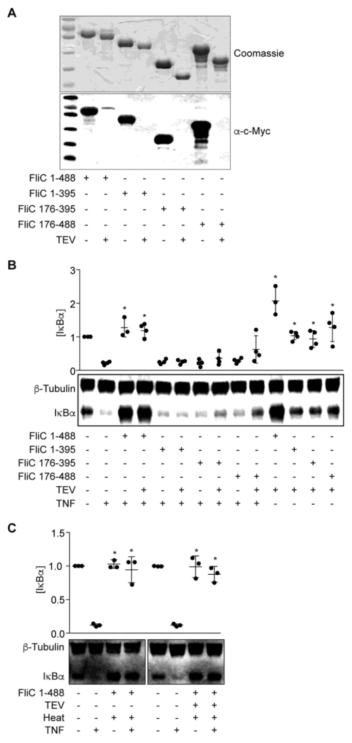
Figure 3.
FliC truncations are inactive. (A) Purified FliC truncations +/− TEV protease treatment were resolved using 10% SDS-PAGE and analyzed by Coomassie blue staining (top) and Western blotting (bottom); (B) IκBα immunoreactivity after incubating HCT-8 cells with FliC truncations (1 µg) for 1.5 h followed by TNF stimulation (20 ng/mL, 20 min); (C) IκBα immunoreactivity after incubating HCT-8 cells with heated (100 °C, 20 min) FliC (1 µg) followed by TNF stimulation (20 ng/mL, 20 min).

