
Figure EV5. ANKRD55 localization and movement along axonemes is consistent with being a component of the IFT‐B particle.
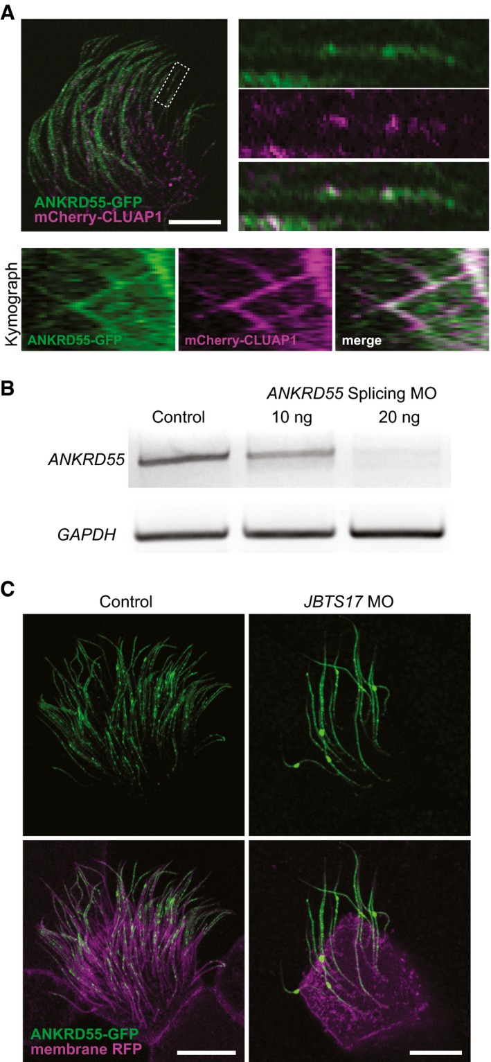
- Two‐color kymograph generated by co‐expression of ANKRD55‐GFP (green) and mCherry‐CLUAP1 (magenta) reveals that ANKRD55 travels along axonemes in association with other IFT proteins. Scale bar: 10 μm. Kymograph is representative out of 22 multi‐ciliated cells.
- RT–PCR demonstrates the efficiency of ANKRD55 MO to disrupt splicing of ANKRD55 mRNA in Xenopus embryos. GAPDH is used as a control.
- Morpholino knockdown of JBTS17, known to specifically affect IFT‐B localization, results in the accumulation of ANKRD55‐GFP in axonemes (green: ANKRD55‐GFP, magenta: membrane RFP). Scale bar: 10 μm. Each image is representative of 18 cells from six different embryos.