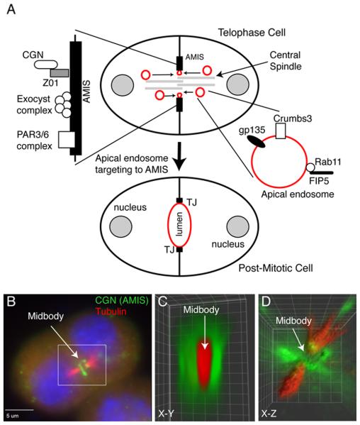
Figure 2. Midbody-dependent recruitment of apical plasma membrane proteins determines the site of nascent apical lumen formation.
(A) Schematic model depicting the role of midbody during lumen formation. Red marks apical endosomes and apical plasma membrane. AMIS stands for apical membrane initiation site.
(B-D) MDCK epithelial cells grown in Matrigel/Collagen matrix were fixed and immunostained with anti-cingulin (CGN; AMIS marker, antibody generated in the Prekeris lab) and anti-acetylated microtubule (central spindle marker) antibodies. Panel B show a single image plane. Panels C and D show 3D rendering based on the entire Z-stack (reproduced from [(Li, Mangan, et al., 2014a)] with permission from EMBO Reports).

