
Figure 6. Flow perturbation promotes neutrophil recruitment through TLR2.
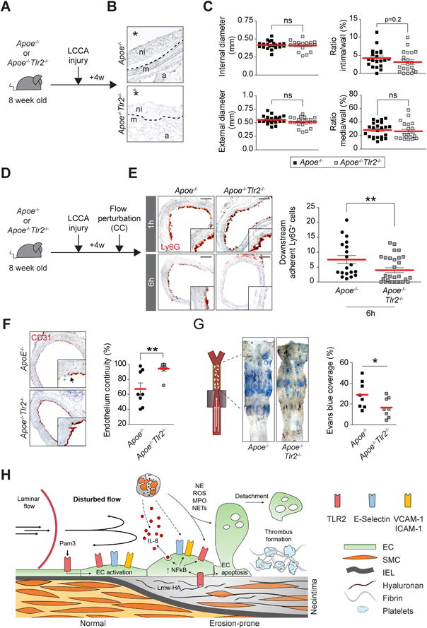
(A) Experimental timeline. (B) H&E staining shows neointima formation after LCCA injury in Apoe−/− and Apoe−/−Tlr2−/− mice. ni: neointima, m: media, a: adventitia. The star indicates the lumen. (C) Morphometric analysis showing measurement of internal (top, left) and external diameters (bottom, left), and the ratio intima/wall (top, right) and media/wall (bottom, right). (D) Experimental protocol involving LCCA injury followed by flow perturbation (CC) in Apoe−/− and Apoe−/−Tlr2−/− mice. (E) Ly6G immunohistochemistry staining of LCCA cross-sections showing the recruitment of neutrophils 1 or 6h after flow perturbation in Apoe−/− and Apoe−/−Tlr2−/− mice. The graph shows the semi-quantitative assessment of adherent neutrophils to the intima after 6h of flow perturbation. Scale bar: 60 μm (F) CD31 immunohistochemistry staining shows endothelium. The graph (right) shows assessment of endothelial continuity (G) LCCA isolated from Apoe−/− and Apoe−/−Tlr2−/− mice were probed for endothelial permeability using Evans blue intravital staining after 6h of flow perturbation. (H) Summary diagram of the main findings of this study. ROS: Reactive oxygen species. MPO: Myeloperoxidase, NETs: Neutrophils extracellular traps, Lmw-HA: Low molecular weight-hyaluronan. IEL: Internal elastic lamina. Data are expressed as mean ± s.e.m. *p<0.05, **p<0.01. Mann–Whitney U test.