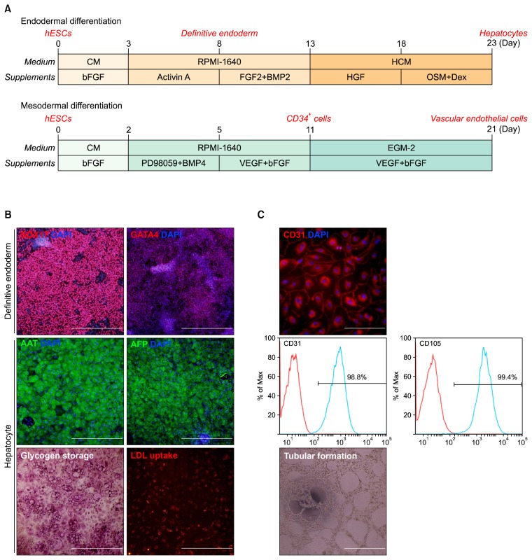
Fig. 1.
Differentiation of hESCs. (A) Overall scheme for the differentiation of hESCs into hepatocytes or vascular endothelial cells. (B) Characterization of endodermal lineage cells derived from hESCs. Immunocytochemistry of SOX17 and GATA4 (up), AAT and AFP (middle), and PAS staining and LDL uptake assay (lower). AAT, anti-α trypsin; AFP, α-fetoprotein; LDL, low-density lipoprotein. Scale bar, 500 μm. (C) Characterization of mesodermal lineage cells derived from hESCs; Immunocytochemistry of CD31 (upper), FACS analysis of vascular endothelial cells (middle), and tubular formation of vascular endothelial cells (lower). Scale bar, 500 μm.

