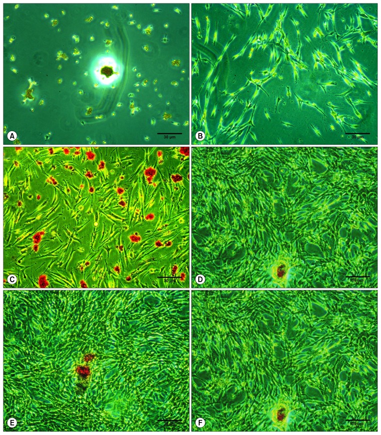
Fig. 4.
Mineralized deposit identified by Alizarin Red S staining in cells grown in osteogenic medium at the 7th day. Alizarin Red S staining of hDPSCs as viewed under the inverted light microscope at 10× magnification with 10% FBS (0.004±0.1095; A), osteodifferentiation media only (0.006±0.1273; B), 1% PRF exudate (0.006±0.1913; C), 5% PRF exudate (0.1273±0.006; D), 10% PRF exudate (0.1290±0.017; E) and 20% PRF exudate (0.1280±0.002; F), Scale bar=30 μm.

