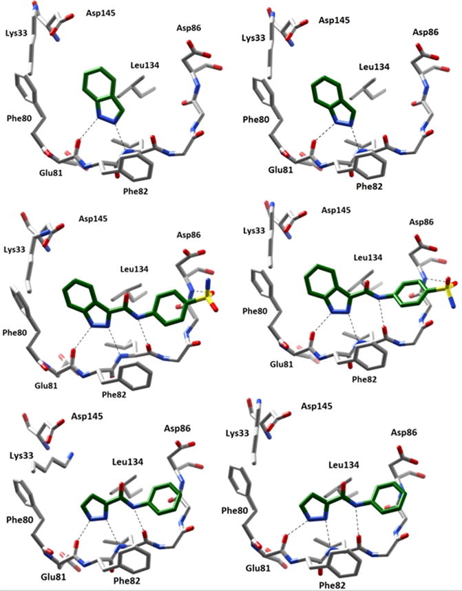
Figure 2.

Comparison of crystalline and docked poses (left and right, respectively) of various CDK2 inhibitors. Dashed lines indicate H-bonding interactions between the ligand and receptor. The docking poses were found to closely resemble those suggested by crystal structures. Top: Compound 1 (PDB: 2VTA16). Middle: Compound 3 (PDB: 2VTI16). Bottom: Compound 4 (PDB: 2VTL16).
