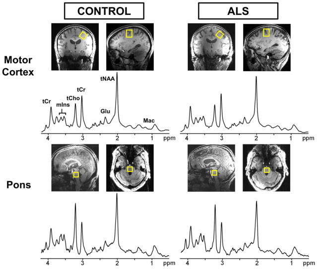
Fig. 1.
Localized proton spectra obtained from the motor cortex (top) and pons (bottom) at 7 T using semi-LASER (TE = 26 ms, TR = 5 s, 64 averages). A 2.2 × 2.2 × 2.2 cm3 voxel (shown on T1-weighted images) was selected in the motor cortex and angulated parallel to the slope of the dural surface in the coronal orientation. A 1.6 × 1.6 × 1.6 cm3 voxel was selected to cover nearly the entire pons region. The spectra are shown with 1-Hz exponential and 5-Hz Gaussian weighting. Left: healthy control (68 year-old female); right: subject with ALS (64 year-old female)

