Figure 3.

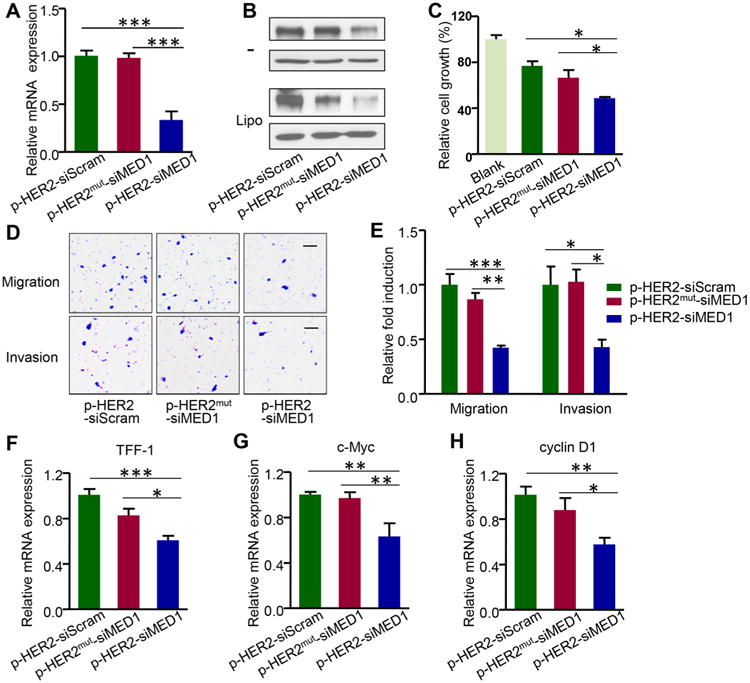
pRNA–HER2apt–siMED1 nanoparticles silenced MED1 expression and inhibited the cell growth and metastatic capabilities of HER2-positive breast cancer cells in vitro. (A) BT474 cells were incubated with 10 μg/mL control and pRNA–HER2apt–siMED1 nanoparticles for 48 h, and the MED1 mRNA level was determined by real-time PCR. (B) BT474 cells were incubated directly with (as indicated by –) or transfected with indicated pRNAs using lipofectamine 2000. At 48 h post treatment, MED1 protein levels were determined by Western blotting. (C–E) BT474 cells were treated with 10 μg/mL pRNA nanoparticles for 48 h and assayed for cell viability by MTT assay (C). Cells were treated as above and seeded for migration (D) and invasion (E) transwell assays. Scale bar: 50 μm. (F–H) After pRNA treatment for 48 h, the mRNA levels of ERα target genes TFF-1 (F), c-Myc (G), and cyclin D1 (H) in BT474 cells were determined by realtime PCR.
